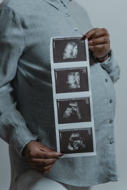
NBME exams are critical assessments for medical students, evaluating clinical knowledge and problem-solving skills. High-yield images in these exams, such as brain scans and pathological findings, are essential for accurate diagnoses and understanding complex conditions, making them a cornerstone of effective preparation and medical education.
Overview of NBME Exams
NBME exams are standardized assessments designed to evaluate medical students’ knowledge and clinical reasoning skills. These exams include a wide range of topics, such as neurological imaging, cardiothoracic pathologies, and abdominal abnormalities. High-yield images, such as brain scans and lung X-rays, are frequently incorporated to test diagnostic accuracy and understanding of complex conditions. The exams are divided into multiple blocks, each focusing on specific medical disciplines. Images are often accompanied by clinical vignettes, requiring students to interpret findings and make informed decisions. These exams are critical for assessing readiness for real-world patient care and are widely used as preparatory tools for licensing examinations like the USMLE.
The Role of High-Yield Images in Exam Preparation
High-yield images play a pivotal role in NBME exam preparation by aiding students in recognizing patterns and diagnosing conditions. These images, often paired with clinical vignettes, focus on commonly tested pathologies, such as brain lesions or pulmonary abnormalities. They enhance visual learning, improving diagnostic accuracy and clinical reasoning; Students rely on these images to familiarize themselves with typical exam content, ensuring they can interpret findings under timed conditions. Access to these images is crucial, as they bridge the gap between theoretical knowledge and practical application, making them indispensable for effective preparation and success in high-stakes medical assessments.
Categories of High-Yield Images in NBME Exams
High-yield images in NBME exams include neurological, cardiothoracic, abdominal pathologies, and other medical imaging, focusing on commonly tested conditions and diagnostic findings.
Neurological and Brain Imaging
Neurological and brain imaging in NBME exams often includes high-yield cases like brain biopsies, metastases, and paraneoplastic syndromes. These images are crucial for diagnosing conditions such as tumors, strokes, and demyelinating diseases. For example, a brain MRI showing metastatic lesions from renal cell carcinoma or paraneoplastic syndromes like polycythemia and hypercalcemia is commonly tested. Such images help assess a student’s ability to correlate radiological findings with clinical presentations, such as neurological deficits or systemic symptoms. These cases emphasize the importance of understanding pathological processes and their imaging manifestations, making them a key focus area for exam preparation and clinical practice.
Cardiothoracic and Abdominal Pathologies
Cardiothoracic and abdominal pathologies are prominently featured in NBME images, covering conditions like pneumonia, aneurysms, and gallstones. These high-yield images often depict chest X-rays showing consolidation patterns or abdominal ultrasounds revealing renal calculi. Such cases test the ability to interpret radiographic findings and correlate them with clinical symptoms, such as chest pain or jaundice. For example, identifying a thoracic aortic aneurysm on a CT scan or recognizing gallbladder wall thickening in ultrasound images is critical. These pathologies are frequently encountered in clinical practice, making them essential for mastery in NBME exams and real-world patient care.

Other High-Yield Medical Images
Other high-yield medical images in NBME exams include those depicting infectious diseases, genetic disorders, and congenital anomalies. For instance, images of Toxoplasma gondii cysts in the brain or cleft lip and palate examples illustrate key pathological and clinical findings. These visuals aid in recognizing patterns and correlations between radiographic abnormalities and clinical presentations, such as paraneoplastic syndromes or metabolic bone diseases. Additionally, images of skin lesions, musculoskeletal abnormalities, and hematological conditions are frequently tested, requiring students to link visual cues with diagnostic criteria. Mastery of these diverse images enhances diagnostic acumen, preparing students for both exam success and real-world patient care.

Sources for Accessing NBME Images
Key sources include the NBME Media Library, shared Anki decks, and medical textbooks like First Aid for the USMLE Step 1, offering high-yield images for exam preparation.
NBME Media Library and Online Repositories
The NBME Media Library serves as a centralized repository for images, videos, and other media artifacts, providing access to high-yield visual content for exam preparation. Students can search the library or upload files to their personal folders for organized study. Online repositories, such as shared Anki decks and medical forums, also offer downloadable PDFs containing NBME images. These resources are invaluable for reviewing clinical vignettes and pathological findings, ensuring familiarity with frequently tested concepts. By leveraging these platforms, learners can enhance their diagnostic skills and improve performance on NBME exams.
Shared Decks and Anki Resources
Shared Anki decks and online resources are invaluable for NBME exam preparation, offering organized access to high-yield images and clinical vignettes. Platforms like AnkiWeb host decks containing NBME images, enabling students to review and master diagnostic findings efficiently. These decks often include tags and spaced repetition systems, optimizing retention of key concepts. Many decks, such as “NBME1-24 images” or “HIGH YIELD IMAGES NBME 25-30,” cover a wide range of topics, from neurological pathologies to abdominal abnormalities. By leveraging these resources, learners can enhance their visual recognition and diagnostic skills, ensuring they are well-prepared for the challenges of NBME exams.
Medical Textbooks and Study Guides
Medical textbooks and study guides are indispensable resources for NBME exam preparation, offering comprehensive collections of high-yield images and clinical vignettes. Textbooks like First Aid for the USMLE Step 1 include over 1,000 color images, focusing on diverse patient demographics and high-yield concepts. These resources often summarize key findings from past NBME exams, such as brain lesions, lung pathologies, and abdominal abnormalities. Many guides also provide concise descriptions of imaging findings, linking them to clinical scenarios and diagnoses. By integrating these materials into study routines, students can enhance their ability to recognize patterns and interpret images accurately, ensuring a strong foundation for success on NBME exams.

How to Effectively Use NBME Images for Preparation
Integrate NBME images into study routines by practicing timed sessions and using active recall. Focus on interpreting clinical vignettes and correlating findings with diagnoses to enhance understanding and accuracy.
Integrating Images into Study Routines
Effectively incorporating NBME images into study routines involves organizing and reviewing them systematically. Start by categorizing images based on body systems or pathologies, such as neurological or cardiothoracic cases. Use active recall by identifying abnormalities without immediate answers. Create flashcards with images on one side and key diagnoses or concepts on the other. Incorporate these into Anki decks for spaced repetition. Dedicate timed sessions to mimic exam conditions, focusing on image interpretation and correlation with clinical vignettes. Regularly review and annotate images, linking them to relevant textbook chapters or online resources. This structured approach enhances visual recognition and diagnostic accuracy, ensuring readiness for high-stakes exams. Consistency and interactivity are key to maximizing learning outcomes.
Timed Practice and Review Strategies
Timed practice is crucial for mastering NBME images, as it simulates exam conditions and enhances time management. Set a timer for 60-90 minutes per session, mirroring the actual test format. Prioritize high-yield images, focusing on common pathologies like brain lesions or pulmonary abnormalities. After each session, review incorrect answers to identify knowledge gaps. Use active recall by attempting to diagnose images without immediate answers. Organize images into categorized decks for systematic review. Incorporate spaced repetition using tools like Anki to reinforce memory retention. Regularly revisit challenging cases to improve diagnostic accuracy. Consistency in timed practice ensures familiarity with image quality and clinical vignettes, optimizing exam performance and confidence. This structured approach is essential for success in high-stakes assessments.
Case Studies and Clinical Vignettes
Case studies and clinical vignettes are invaluable for mastering NBME images, as they provide real-world context and enhance diagnostic skills. These tools present scenarios with patient histories, symptoms, and imaging findings, mimicking actual clinical encounters. By analyzing high-yield images within these narratives, students learn to correlate radiographic findings with clinical presentations, such as brain metastases in a patient with a history of malignancy or pulmonary masses in immunocompromised individuals. Regular review of these cases improves pattern recognition and the ability to prioritize differentials. Integrating case studies into study routines bridges the gap between theoretical knowledge and practical application, ensuring a deeper understanding of complex pathologies and their imaging characteristics.

Future Trends in NBME Imaging and Assessment
Future trends include enhanced image quality, advanced annotations, and improved digital accessibility, enabling more precise and interactive assessments for medical students and professionals.

Advancements in Image Quality and Annotation
Recent advancements in image quality and annotation have significantly enhanced the clarity and educational value of NBME images. High-resolution scans and detailed annotations provide precise diagnostic cues, aiding students in identifying pathologies. Improved color accuracy and contrast ensure that subtle abnormalities are more apparent, mimicking real-world clinical scenarios. Additionally, interactive annotations now offer layered information, allowing learners to explore images at varying depths. These innovations not only improve understanding but also prepare students for the evolving demands of medical practice. Enhanced annotations further standardize testing, ensuring consistency and fairness in assessments. These advancements underscore the importance of visual learning in medical education.
Digital Platforms and Accessibility
Digital platforms have revolutionized access to NBME images, enabling medical students to study efficiently. Online repositories like the NBME Media Library offer searchable databases of high-yield images, while shared decks on Anki and other platforms allow collaborative learning. Mobile accessibility ensures that learners can review images anytime, anywhere. Digital tools also support interactive features, such as zooming and annotation, enhancing the study experience. These platforms foster flexibility and engagement, catering to diverse learning styles. By centralizing resources, digital platforms reduce barriers to accessing high-quality materials, empowering students to prepare effectively for exams. This shift underscores the growing importance of technology in modern medical education.
Mastering NBME images is a cornerstone of exam success. Consistently reviewing high-yield images sharpens diagnostic skills and reinforces clinical knowledge. Incorporate image-based questions into study routines, focusing on annotations and contextual clues. Prioritize active learning over passive review, and seek feedback to identify knowledge gaps. Stay updated with the latest NBME resources and digital platforms for enhanced accessibility. Time management and structured practice are key to excelling. By integrating these strategies, learners can optimize their preparation and confidently approach the NBME exams, ensuring a strong foundation for future medical practice.